
ECG
The ECG procedure may not be difficult to perform but surely requires competence and accuracy. Each electrical current of the heart must be precisely traced as this is what helps in deciding upon the course of care and treatment. Even a slight mistake may result in an inaccurate reading and lead to the wrong medication or treatment procedure.
- Identifying and placing leads correctly on a virtual patient
- Understanding the controls of an ECG machine and steps to generate a report
- Mastering the five-step process of gathering and interpreting ECG data
- Identifying sinus rhythms and analyzing rhythm strips
- Identifying inverted P morphology formations, P wavelengths and their relation to QRS complexes

Developed in alignment with Electrocardiography for Healthcare Professionals (Fifth Edition) by Kathryn Booth and Thomas E. O’Brien.
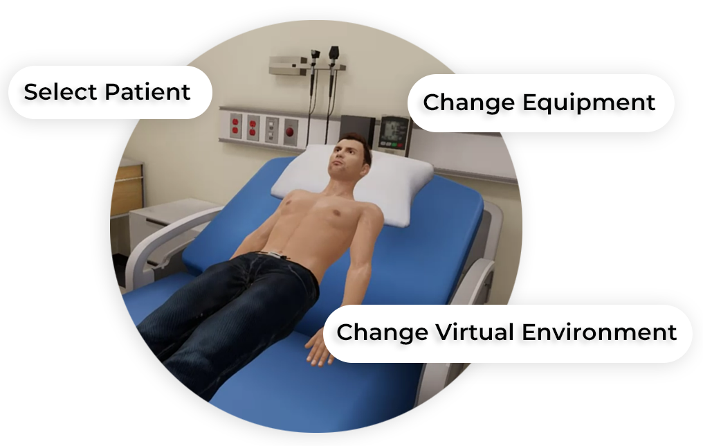
Customize Your Program
Get rid of the editor. Adopt in-VR customization.
MedVR Education is bringing to you in-VR customization that will enable you to customize your procedural simulations by making selections from a range of feature choices.
- Select patient from a diverse background
- Choose preferred virtual environment
- Select equipment used in the procedure
- Modify difficulty level of the assessment mode
- …..many more to come
 Physics-based Interaction
Core Skills Training
Leads and Waveforms
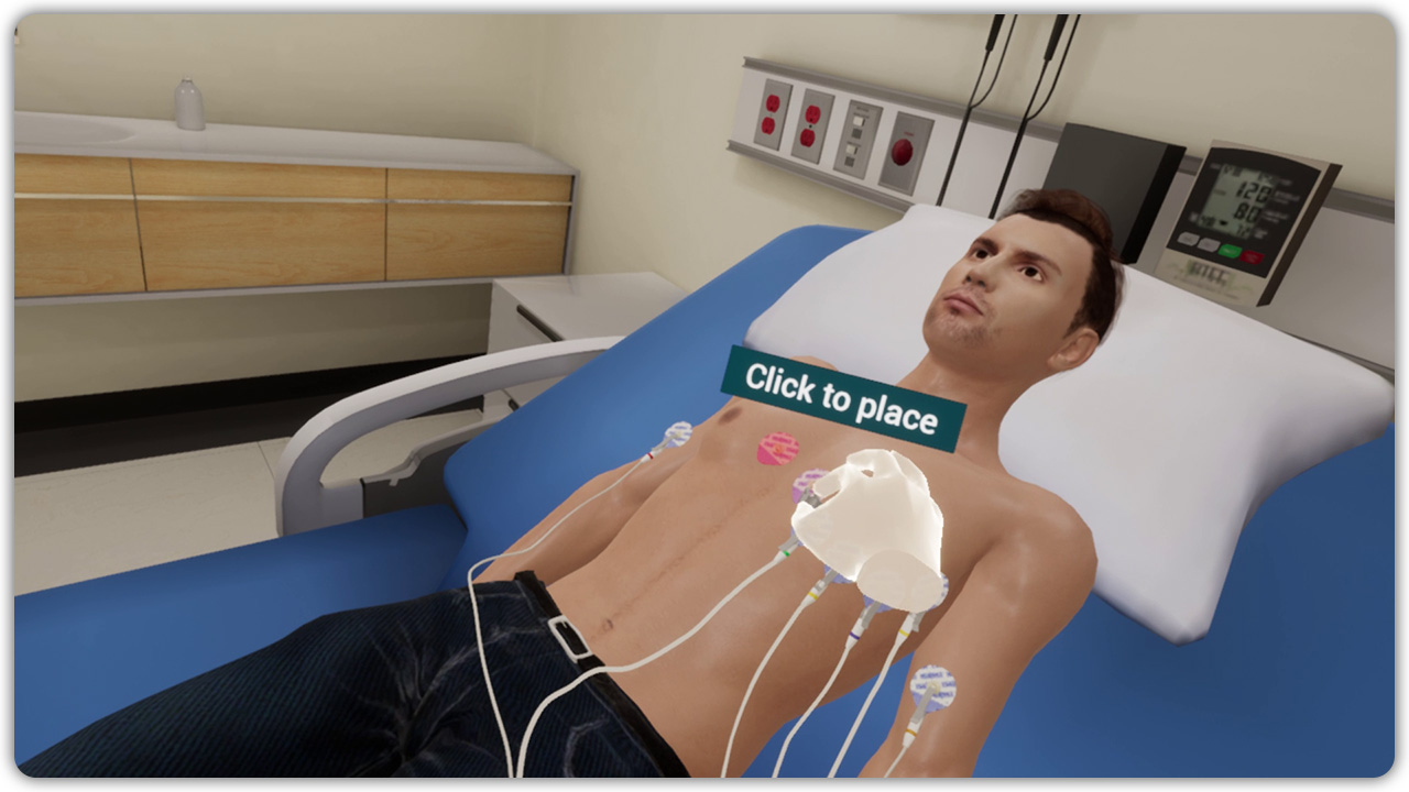
The 12-lead ECG, that records the electrical activities of the heart, makes use of electrodes and lead wires to complete the process. Lead wires are connected to electrodes, small sensors, on one end and to the ECG machine on the other end. The electrodes are placed on the skin to read the electrical activities which are transmitted to the ECG machine through the lead wires. Though named 12-lead ECG, 10 lead wires are used for the purpose. This VR training, helps learners reinforce their knowledge about the 12-lead ECG system and how the electrical activity works across various leads. Theoretical knowledge is applied in a practical scenario containing a virtual patient and various interactive tools. The interactions have been designed to help learners visualize directions of currents produced by various lead combinations.
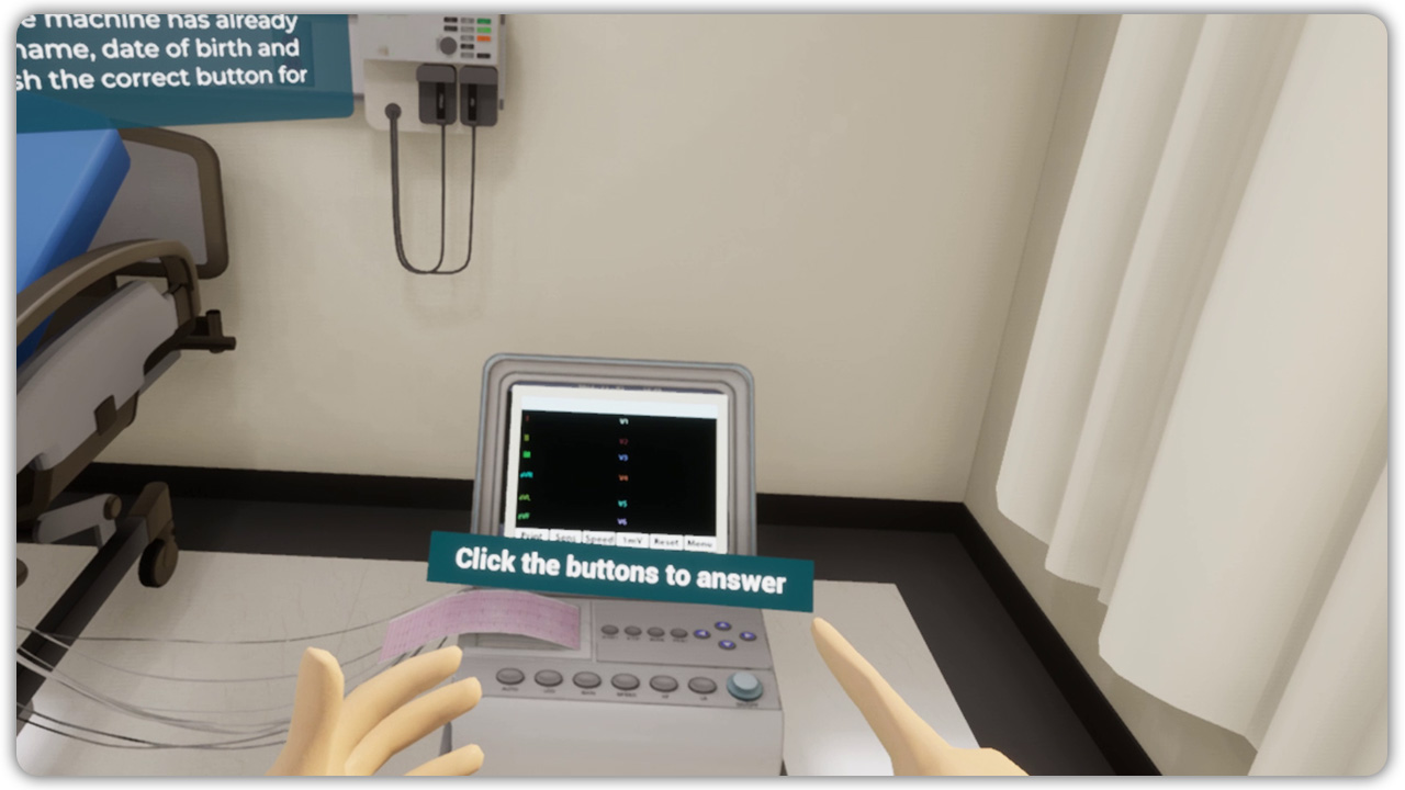
Operating ECG Machine
Speed, gain and artifact filter – the three most important controls on the electrocardiograph. This VR training program is designed to help learners get an insight into the controls on the electrocardiograph. Learners will benefit by enhancing their skills required to operate the ECG machine based on the use case. Interacting with the machine, gaining familiarity with its functions, and performing steps to generate the correct output of the graph will also assist in building confidence. A confident ECG operator can always help put an anxious patient at ease for the procedure, thereby making the process smooth and comfortable. Learners can also gain practical knowledge of the practical implications faced while dealing with a patient.
Performance of an ECG
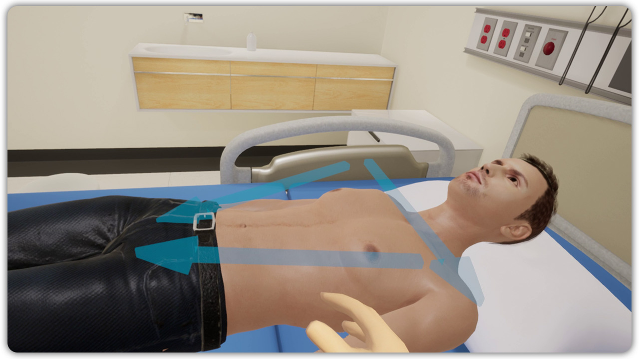
In order to perform a successful ECG procedure, it is important to have prefect knowledge of how to place the electrodes on the chest and limbs as well as understand the significance of the limb leads. ECG readings need to be accurate and precise. The precision of readings gained from this procedure is particularly important when it concerns patients suffering a cardiac crisis. Placing electrodes at the wrong places will produce an incorrect result or no graph in the output. This VR training, with the use of virtual humans, provides learners with an immersive experience to help them practice and perform the lead placements accurately as well as connect the correct lead wires for a successful ECG procedure.
Rhythm Interpretation
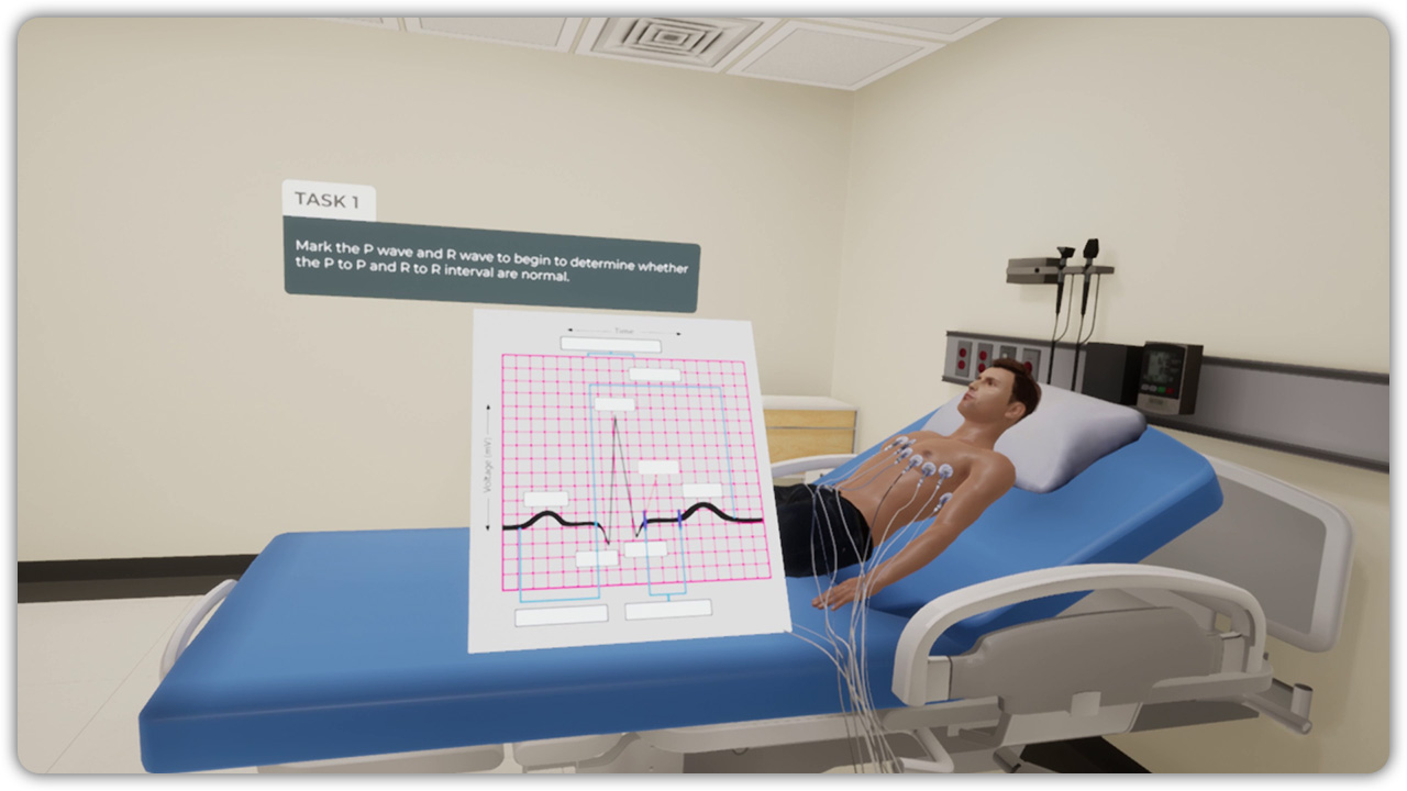
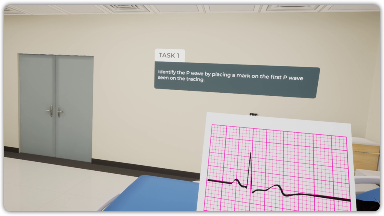
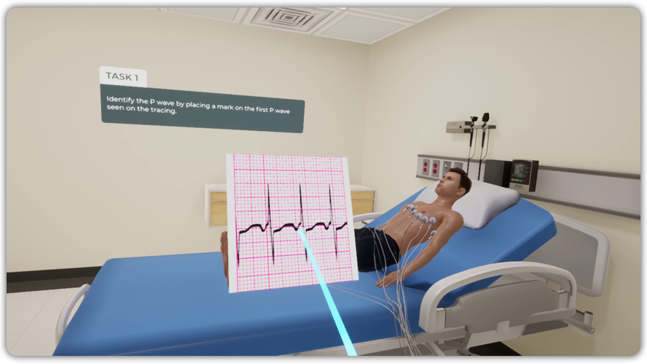
An ECG analysis is a combination of five-steps that are followed to gather data about the five components of the ECG rhythm strip. These components consist of Rhythm (regularity), Rate, P wave morphology (shape), PR interval, QRS duration and morphology. In the immersive environment of this VR training session, learners master the five-step process of gathering data, perform interpretations, and get skilled in rhythm classification. Learners will be presented with an output graph of the ECG and they can select and highlight the correct region on the graph.
Sinus Rhythm
For every patient, a normal sinus rhythm is the desired rhythm. A patient with a normal sinus rhythm should have a normal cardiac output. Cardiac Output (CO) refers to the volume of blood the heart pumps each minute. Stroke volume is the volume of blood ejected by one or both ventricles with each contraction. CO is determined by using the equation Heart Rate (HR) X Stroke Volume (SV). This VR session will help learners become proficient in identifying various sinus rhythms originating from the sinus node by analyzing the rhythm strips. This competency is extremely helpful to improve the analytical skill, interact with the graphs and identify the correct rhythms produced.
Junctional Dysrhythmias
The AV junction includes the atrioventricular (AV) node and the surrounding area, including the bundle of His. When it comes to junctional rhythms it is worth noting that the electrical currents originate from the AV junction and not the SA node. Due to this reason the electrical impulses that result in depolarization of the atria flow in the reverse direction. This backward flowing electrical impulse leads to the formation of an inverted P morphology which is seen in junctional dysrhythmias. This VR session is designed to help learners’ practice and get skilled in identifying these formations, P wavelength and its relation to the QRS complexes.