Tunneled Vascular Access Placement

SIMULATION TYPE
Procedural

SIMULATION DURATION
15 minutes

PATIENT AGE GROUP
Adult

TARGET AUDIENCE
Medical Students, PA, and Physicians

Fifty-eight-year-old Michael Carter is scheduled for a tunneled vascular access catheter placement for long-term dialysis. The procedure requires precision, adherence to aseptic technique, and effective use of imaging to ensure safe and accurate vascular access.
This VR simulation provides healthcare professionals with a realistic opportunity to practice the procedure from start to finish in an immersive, controlled environment. Learners are guided through patient preparation, sterile setup, venous access under ultrasound, wire advancement, tunneling, and post-placement verification.
The module offers both training and assessment modes to support skill acquisition, procedural accuracy, and clinical decision-making in a simulated operating environment.
- Preparing the patient and maintaining a sterile field for vascular access
- Using clinically approved techniques to perform venous access using ultrasound guidance
- Advancing and exchanging guidewires under fluoroscopic visualization
- Accurately creating and using a subcutaneous tunnel for catheter placement
- Testing, securing, and dressing the catheter appropriately
- Confirming final placement and catheter functionality
- Pereira, K., et al. (2015). Vascular access for placement of tunneled dialysis catheters for hemodialysis: A systematic approach and clinical practice algorithm. Clinical Imaging Science, (5), 24.
Customize Your Program
Get rid of the editor. Adopt in-VR customization.
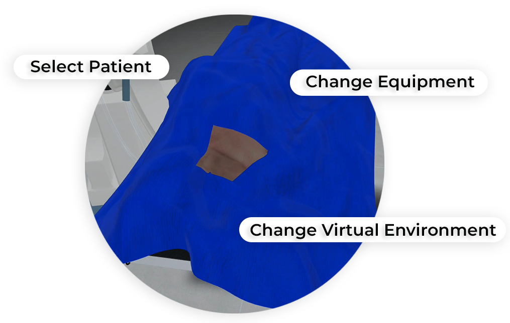
MedVR Education is bringing to you in-VR customization that will enable you to customize your procedural simulations by making selections from a range of feature choices.
- Select patient from a diverse background
- Choose preferred virtual environment
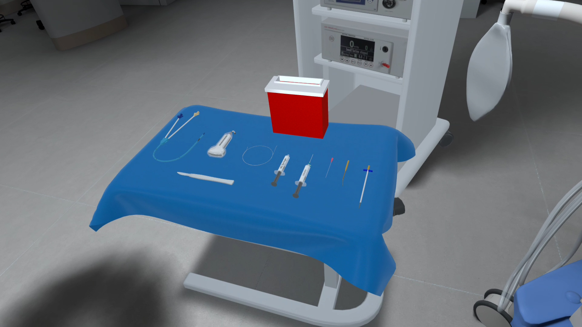
- Select equipment used in the procedure
- Modify difficulty level of the assessment mode
- …..many more to come
 Multi-playerSessions
 Physics-based Interactions
Core Skills Training
Performing Tunneled Vascular Access Placement
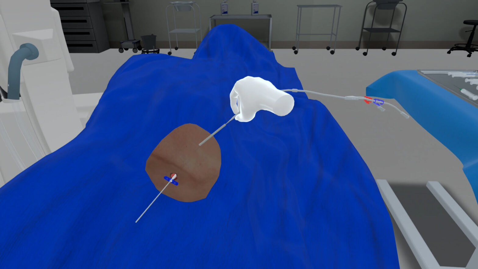
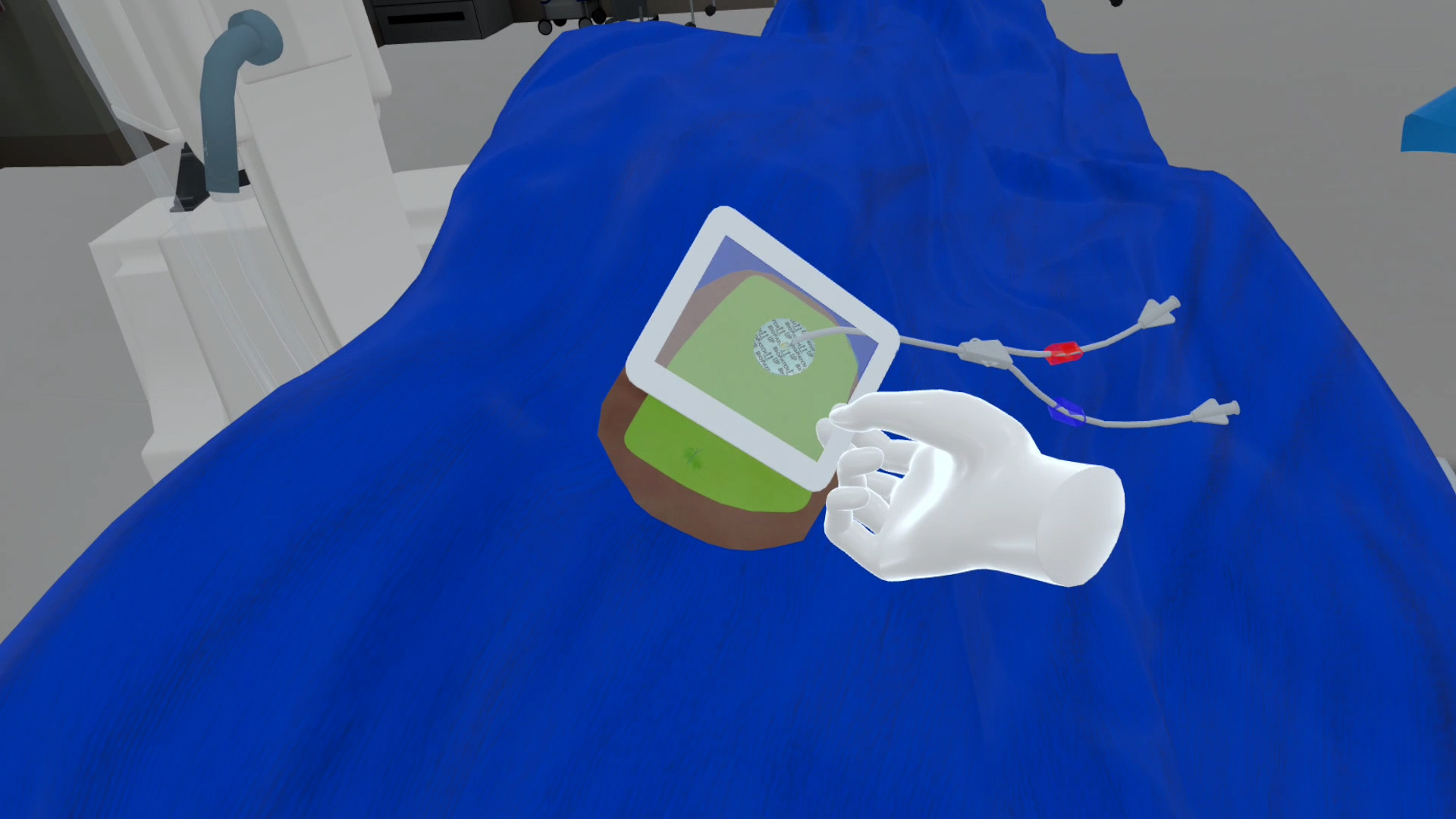
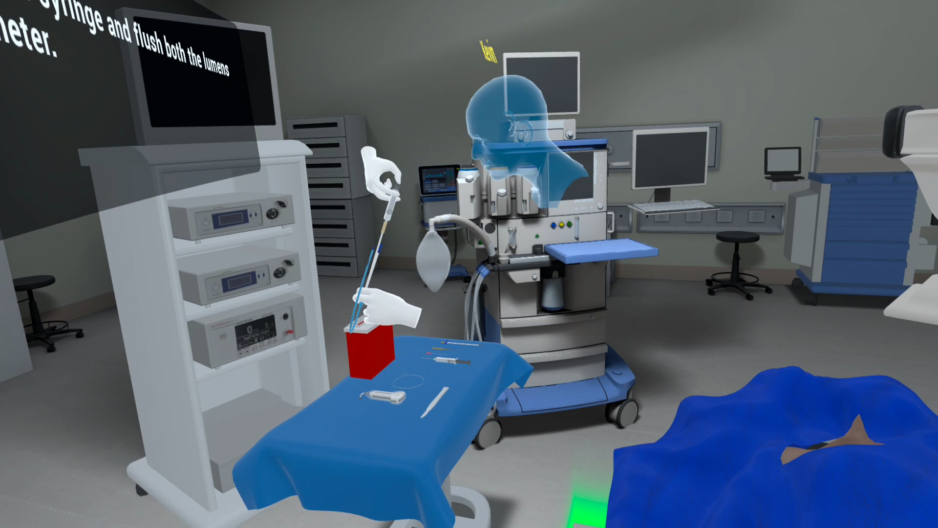
As part of this Tunneled Vascular Placement procedure, the learner begins by verifying patient identity, confirming allergies, and explaining the procedure. After hand hygiene, the patient is prepped and draped in a sterile field. Using a linear ultrasound probe, the learner identifies the right internal jugular vein, administers local anesthesia, and advances the access needle under ultrasound visualization. Once venous entry is confirmed, a guidewire is introduced and advanced under fluoroscopy. Sequential dilators and a peel-away sheath are inserted to prepare the tract. The catheter is tunneled subcutaneously from the chest entry site to the venotomy point and advanced under fluoroscopic guidance to the correct location. Catheter placement is confirmed with fluoroscopy, followed by aspiration and flushing of each port to ensure patency. The site is sutured, dressed with a biopatch, and covered with a sterile dressing to complete the procedure.
Training
With prompts, guidance and affordances learners are hand-held through the process to practice the procedure in a virtual environment with a virtual patient.
- Photorealistic virtual environment
- Physics-based interactions
- Detailed instructions
- Adequate affordances to assist in task completion
Assessment
Test acquired skills to perform the procedures from start to finish without prompts. An incorrect step will take the learner back to the start to start afresh.
- Live scoring
- Instant feedback
- Adequate affordances for efficient performance
- Time tracking to monitor activity completion