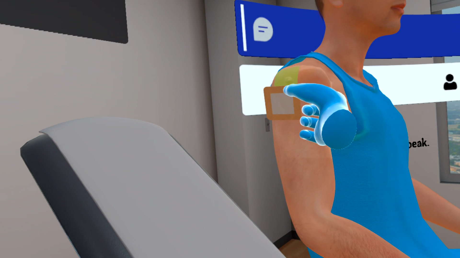
Reduction of Anterior Shoulder Dislocation

SIMULATION TYPE
Procedural

SIMULATION DURATION
20–30 minutes

PATIENT AGE GROUP
Adult (23-Year-Old Male)

TARGET AUDIENCE
Medical Students, Nursing Students, Emergency Medicine Learners

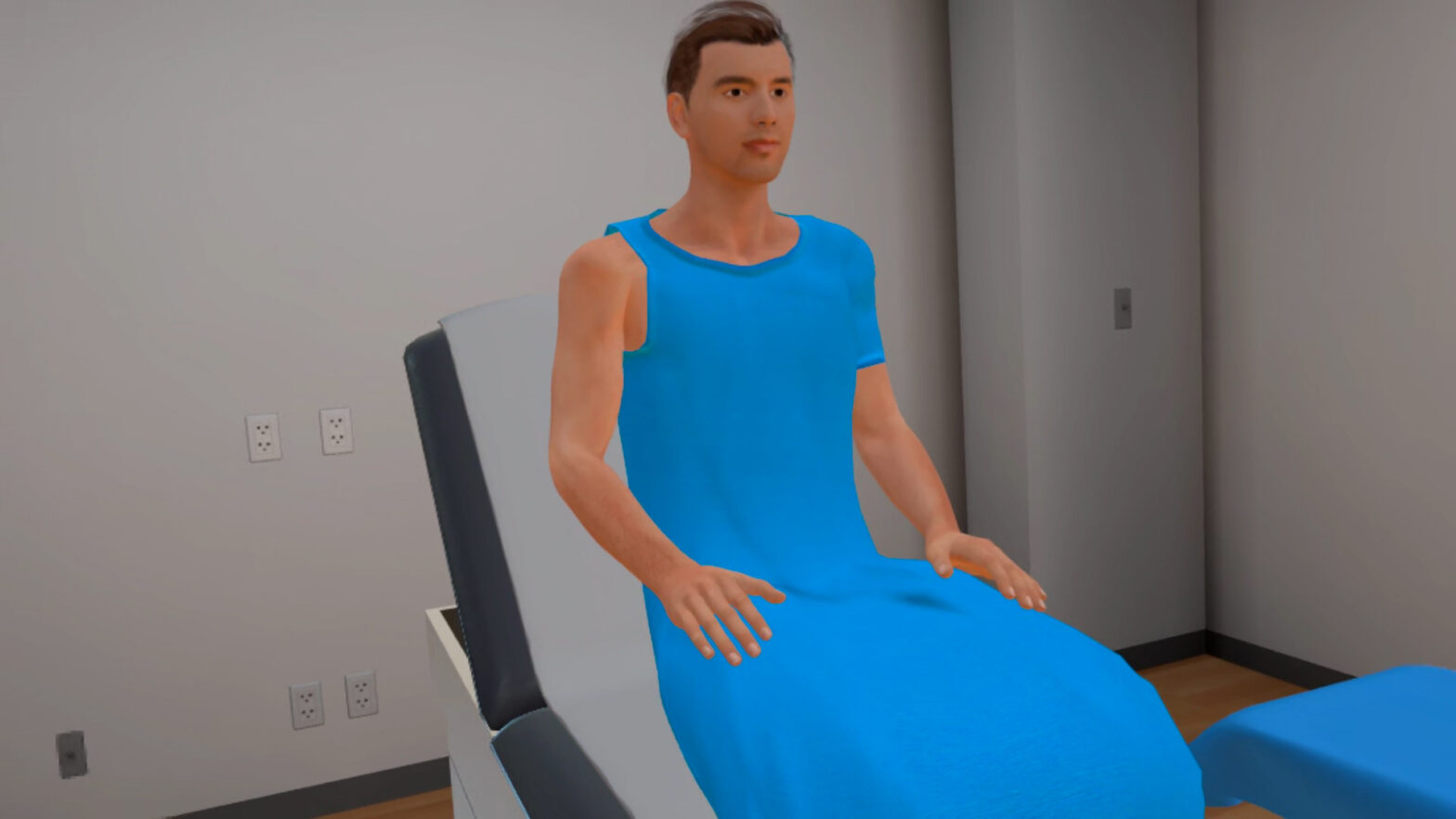
Twenty-three-year-old male presents to the Emergency Department with right shoulder pain and inability to move his right shoulder following a volleyball injury. He reports diving for a ball and landing on his right shoulder. Clinical examination reveals the right arm held slightly abducted and externally rotated, with a prominent acromion and findings consistent with an anterior shoulder dislocation.
This VR simulation provides learners with a realistic, immersive environment to practice the assessment and reduction of an anterior shoulder dislocation in a controlled Emergency Department setting. Learners progress through patient assessment, neurovascular examination, diagnostic imaging, analgesia, intra-articular lidocaine administration, shoulder reduction, post-reduction assessment, immobilization, confirmatory imaging, and discharge planning while following clinical best practices.
The module includes training and assessment modes to reinforce correct procedural sequencing, patient communication, clinical examination, reduction technique, neurovascular assessment, equipment handling, and post-procedure care.
- Assess a patient presenting with suspected anterior shoulder dislocation
- Recognize the role of appropriate diagnostic imaging before and after reduction
- Prepare for and perform an intra-articular lidocaine injection
- Maintain effective patient communication and comfort throughout the procedure
- Reassess motor, sensory, and distal vascular function following reduction
- Complete appropriate post-procedure care and discharge planning
- Ufberg, J. W., Vilke, G. M., Chan, T. C., & Harrigan, R. A. (2004). Anterior shoulder dislocations: Beyond traction-countertraction. The Journal of Emergency Medicine.
Customize Your Program
MedVR Education works with your organization to tailor the anterior shoulder dislocation simulation to your educational goals, clinical workflows, and learner experience. Customizations are developed in collaboration with your team to meet your specific training needs.
- Customize patient presentation and injury scenarios
- Configure Emergency Department room layouts
- Adjust procedural difficulty and assessment criteria
- Support preferred shoulder reduction techniques
- Additional customizations available upon request to align with your program objectives
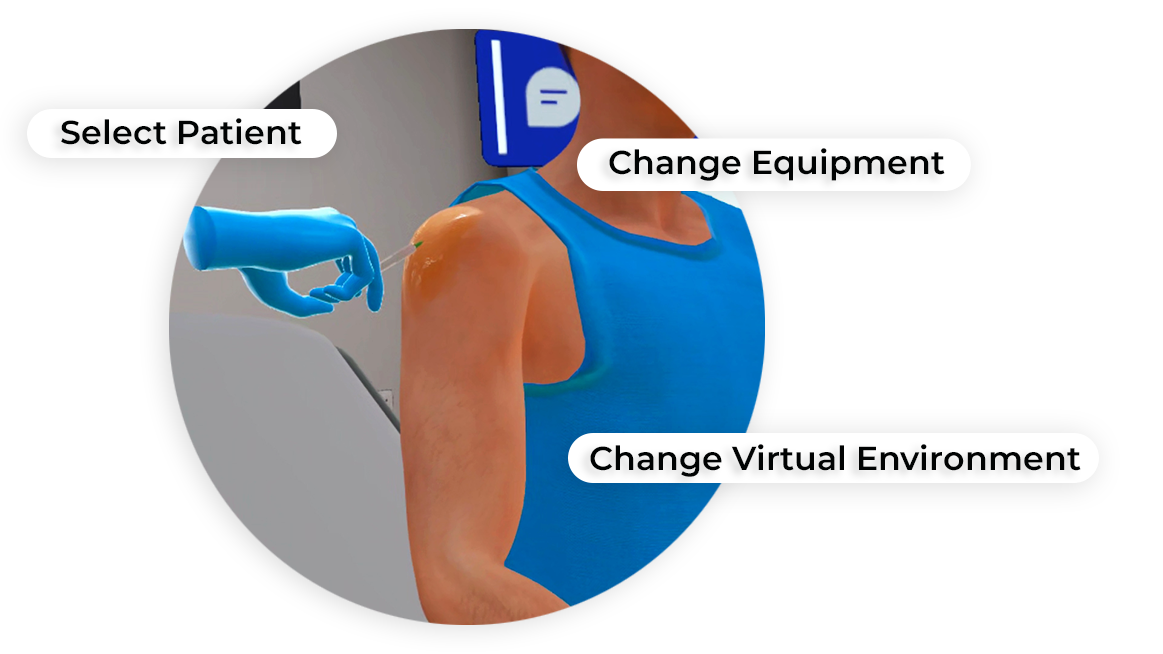
 AI Patient Assessment
 Natural Language Processing
 Multi-playerSessions
 Physics-based Interactions
Core Skills Training
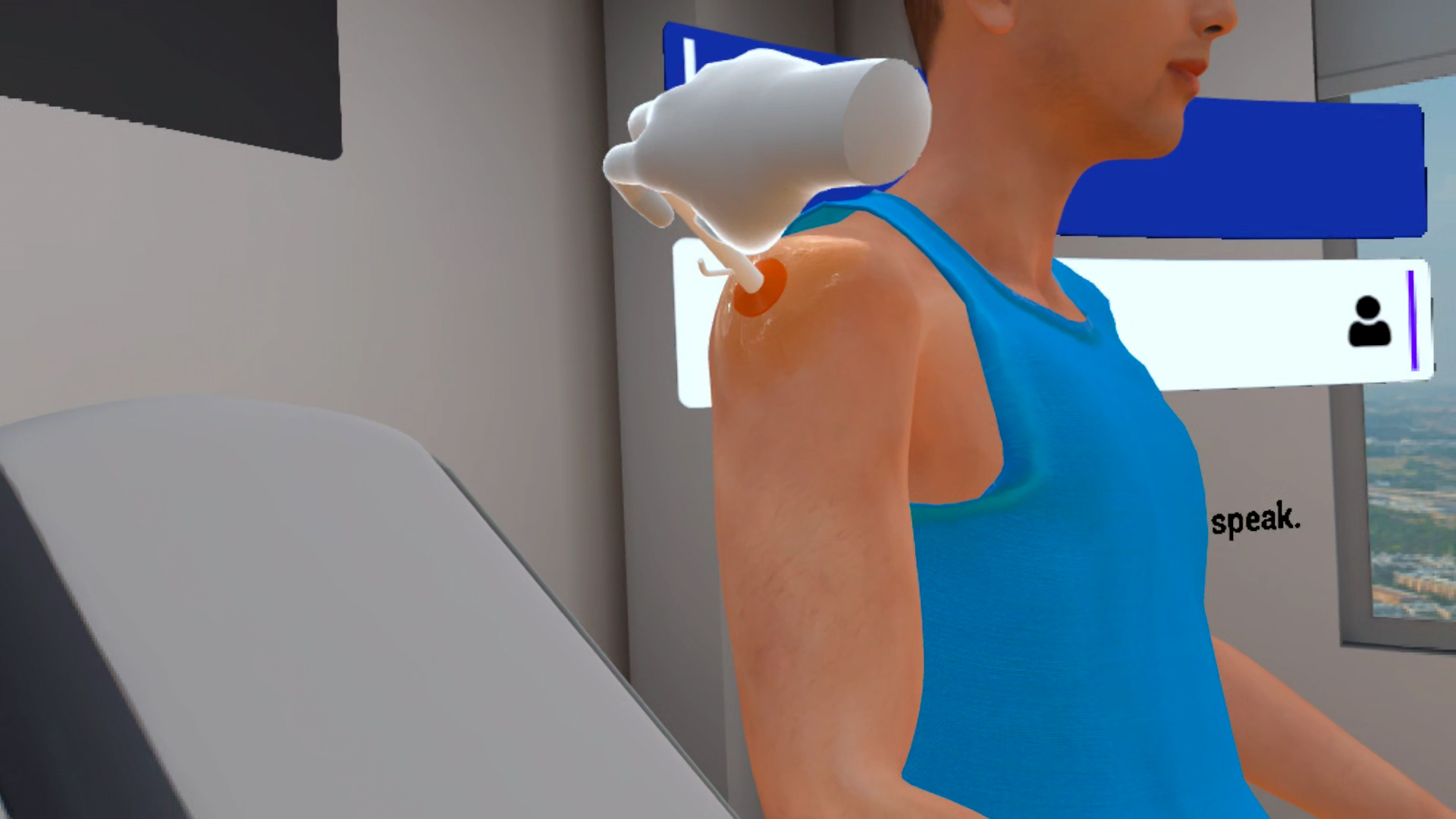
Performing Reduction of Anterior Shoulder Dislocation
The learner begins by assessing the patient and affected shoulder, including arm position, bony tenderness, joint range of motion, motor and sensory function of the median, ulnar, radial, and axillary nerves, and distal pulses. The learner prepares and administers intra-articular lidocaine using proper equipment, injection-site identification, chlorhexidine skin preparation, sterile technique, and adequate anesthetic time.The learner then performs shoulder reduction using an appropriate technique, including external rotation, FARES, or Cunningham. For external rotation, the learner positions the patient upright, flexes the elbow to 90°, slowly externally rotates the arm, allows muscle relaxation, then abducts with downward traction until reduction occurs.
Training
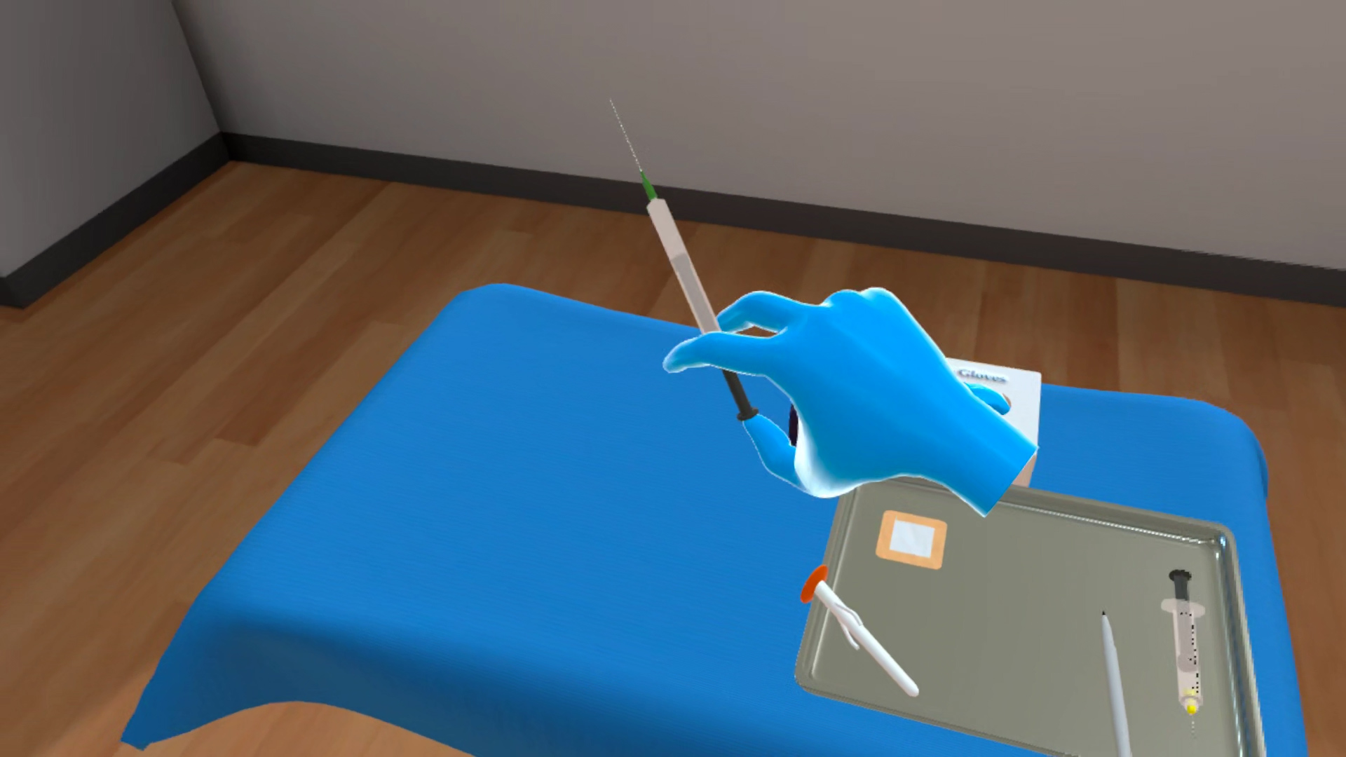
With prompts, guidance, and affordances, learners are guided step-by-step through the assessment and reduction of an anterior shoulder dislocation in a virtual Emergency Department environment.
- Photorealistic Emergency Department environment
- Physics-based interaction with the patient and clinical equipment
- Guided instructions and visual cues
- Affordances supporting patient assessment and neurovascular examination
- Guided practice with intra-articular lidocaine administration
- Support for post-reduction assessment and immobilization
Assessment
Learners demonstrate independent performance of the procedure from start to finish without prompts. Incorrect steps require the learner to correct the procedure and complete the task appropriately.
- Live scoring
- Immediate feedback
- Sequence and technique validation
- Neurovascular assessment validation
- Time tracking for task completion